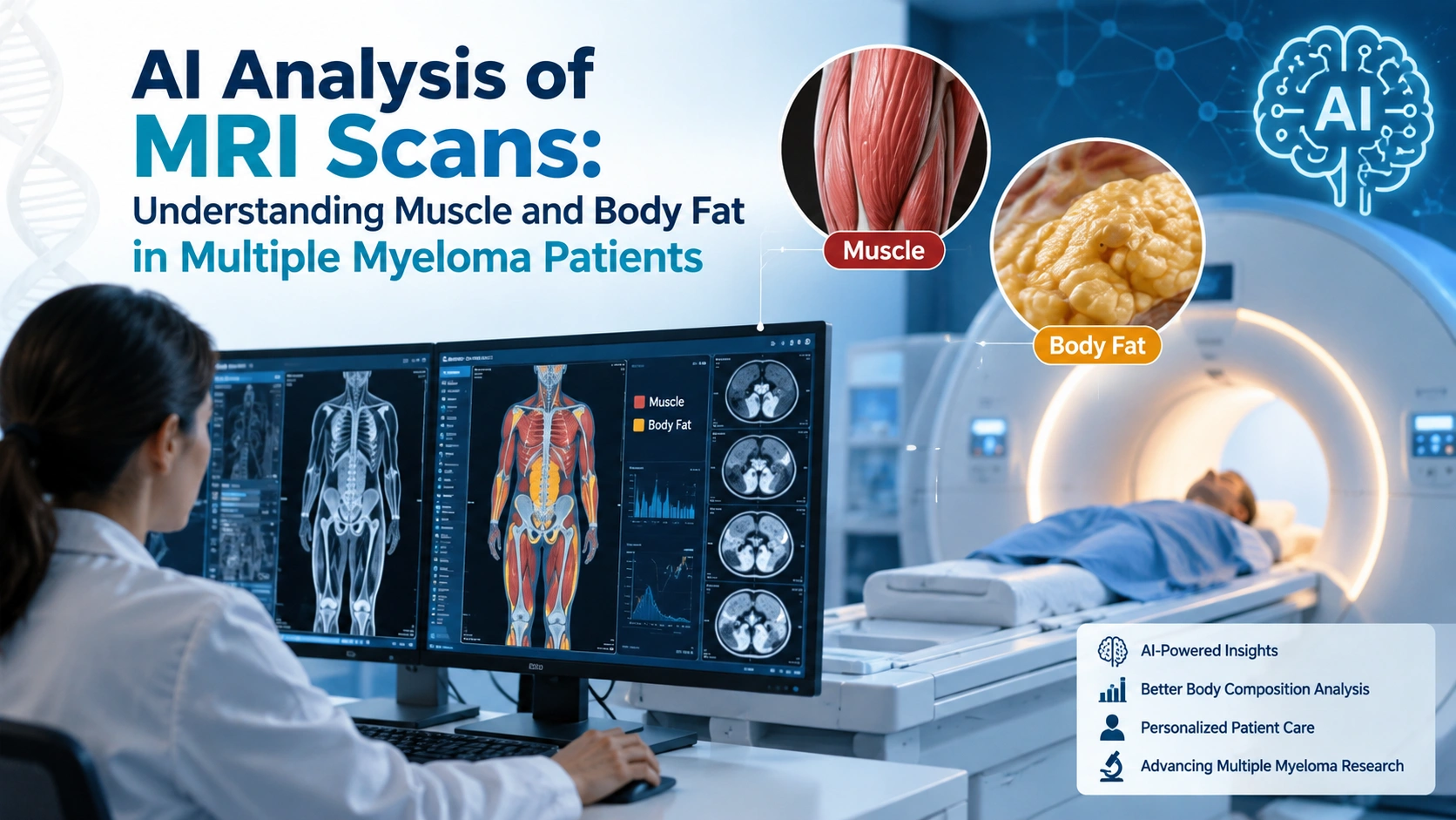
A routine MRI scan is usually ordered to look at disease activity. But could the same scan reveal something else about a patient’s health—such as how much muscle and body fat they have?
New research suggests it may be possible. Scientists at The Royal Marsden NHS Foundation Trust and The Institute of Cancer Research developed an artificial-intelligence pipeline that can analyze routinely acquired whole-body MRI scans in people with multiple myeloma and automatically measure body-composition features.
The findings are promising, but they should be understood as research rather than a new standalone diagnostic or treatment tool.
What Is Multiple Myeloma?
Multiple myeloma is a blood cancer involving abnormal plasma cells in the bone marrow. Treatment can involve multiple lines of therapy over time, making a patient’s overall physical condition and resilience important considerations.
Multiple myeloma can affect people differently, and researchers are increasingly studying factors beyond the cancer itself—including muscle and fat composition—to better understand outcomes.
Why Does Body Composition Matter?
Body composition describes the amount and distribution of tissues such as skeletal muscle and body fat. It can provide information that body weight or BMI alone may not capture.
For example, two people can have the same BMI but very different amounts of muscle and visceral fat. Previous research in multiple myeloma has found associations between measures such as sarcopenia, muscle quality and adipose tissue and clinical outcomes, although results have not always been consistent.
How Can AI Analyze an MRI?
AI can be trained to identify and segment different tissues in medical images automatically. In the new MRI study, a deep-learning pipeline was used to extract quantitative body-composition measurements from routinely acquired whole-body MRI scans. (PubMed)
The process can be viewed simply:
- A patient undergoes a clinically indicated whole-body MRI.
- AI analyzes the images.
- The system identifies relevant tissues.
- Muscle and fat measurements are calculated.
- Researchers can compare these measurements with clinical outcomes.
This approach is important because it may allow researchers to obtain additional information from scans that are already being performed.
What Did the New MRI Study Find?
The researchers observed changes in muscle and fat during treatment, and some baseline body-composition measures were associated with progression-free survival.
The study found a decrease in abdominal skeletal muscle during treatment, alongside temporary increases in abdominal subcutaneous and visceral adipose tissue. Greater abdominal skeletal muscle reserves and greater subcutaneous adipose tissue at baseline were associated with better progression-free survival. In contrast, increasing visceral adipose tissue over time was associated with poorer progression-free survival.
These are associations, not proof that changing muscle or fat directly causes better or worse cancer outcomes.
What Is Sarcopenia?
Sarcopenia refers to reduced muscle mass, muscle quality or strength. It can be particularly relevant when studying older adults and people undergoing demanding cancer treatments.
Earlier multiple myeloma research has also explored AI-assisted identification of sarcopenia from CT imaging. One study found that CT-identified sarcopenia was independently associated with survival in newly diagnosed multiple myeloma.
However, other studies have reported different findings, showing why larger prospective studies are needed.
MRI vs CT for Body Composition
| Feature | MRI | CT |
|---|---|---|
| Radiation | No ionizing radiation | Uses ionizing radiation |
| Tissue information | Strong soft-tissue contrast | Strong anatomical and tissue-density information |
| AI analysis | Can automate tissue segmentation | Can automate tissue segmentation |
| Multiple myeloma research | Emerging opportunity | More established research base |
| Routine use | Depends on clinical indication | Often available from existing scans |
CT-based AI body-composition analysis has already been investigated in multiple myeloma. A 2025 study of 91 patients found that AI-derived body-composition measures could contribute to patient stratification, although the authors emphasized that more research is needed.
Why Is AI Useful Here?
AI can turn visual information from medical images into measurable data. Instead of relying only on a clinician’s visual assessment, automated segmentation can quantify muscle, subcutaneous fat, visceral fat and other tissues.
This could potentially make body-composition analysis more scalable and reproducible across large datasets.
Could Routine MRI Become a New Risk Tool?
Possibly, but the evidence is still developing. The 2026 MRI study reported a concordance index of 0.725 for its body-composition-based risk-stratification approach, suggesting potential predictive value.
However, this does not mean MRI-based AI can currently predict an individual patient’s outcome with certainty.
Clinical decisions still depend on factors such as disease characteristics, laboratory findings, treatment response, genetics and overall health.
What Could This Mean for Patients?
If validated in larger studies, opportunistic body-composition analysis could eventually help clinicians identify patients who may need closer attention to nutritional status, physical function or treatment-related changes.
For example, repeated scans might reveal loss of muscle or changes in fat distribution that are not obvious from body weight alone.
The goal would not be to replace clinical assessment, but potentially to add another measurable layer of information.
What Are the Limitations?
The research is promising, but several questions remain:
- The MRI findings need validation in larger and diverse patient populations.
- Association does not prove causation.
- AI algorithms need testing across different scanners and institutions.
- Body composition is only one part of multiple myeloma prognosis.
- Research findings should not be used to make individual treatment decisions without medical guidance.
Other body-composition studies in multiple myeloma have produced mixed results, reinforcing the need for careful interpretation.
The Future of AI and Multiple Myeloma Imaging
The emerging idea is simple: a medical scan may contain more information than the original clinical question it was ordered to answer.
AI could help researchers extract that additional information efficiently, turning routine whole-body MRI into a potential source of quantitative data about muscle and fat.
For multiple myeloma, this may eventually support more personalized approaches to risk assessment and supportive care—but further prospective research is needed before such methods become routine clinical practice.
Frequently Asked Questions
Can AI analyze MRI scans in multiple myeloma?
Yes. Research has demonstrated that deep-learning systems can automatically extract body-composition measurements from routinely acquired whole-body MRI scans in multiple myeloma patients.
Can MRI show muscle and body fat?
Yes. MRI provides detailed soft-tissue information, and AI can be used to identify and quantify different tissues within whole-body scans.
Does low muscle mass mean worse multiple myeloma?
Not necessarily. Research has found associations between muscle-related measures and outcomes, but findings vary between studies. Low muscle mass should not be interpreted as an individual prognosis.
Can AI predict multiple myeloma outcomes?
AI-based body-composition analysis shows potential for risk stratification, but it is not currently a standalone method for predicting an individual patient’s outcome.
Is AI replacing doctors in MRI analysis?
No. AI research in this area is intended to assist image analysis and extract quantitative information. Clinical interpretation and treatment decisions remain the responsibility of qualified healthcare professionals.
Conclusion
AI analysis of MRI scans could give researchers and clinicians a new way to understand muscle and body fat in multiple myeloma. The latest research suggests that body composition changes during treatment may be linked with progression-free survival, but these findings require further validation.
The bigger opportunity is not simply “AI reading an MRI.” It is turning routine medical images into additional, measurable information that could support more personalized cancer care.